Congenital epidermolysis bullosa diagnosis
Diagnosing congenital epidermolysis bullosa (EB) involves a detailed clinical evaluation, supplemented by advanced laboratory techniques. EB is a group of rare skin disorders characterized by fragile skin that blisters easily. A precise diagnosis not only confirms the presence of the condition but also helps identify the specific subtype, which is crucial for managing the disease, offering genetic counselling, and predicting long-term outcomes. Modern diagnosis of congenital epidermolysis bullosa combines physical examination methods with tests that assess protein expression and underlying genetic mutations, providing a clear understanding of the patient's disease.
How is congenital epidermolysis bullosa diagnosed?
The process of diagnosing EB begins with a clinical evaluation, where clinicians review the patient’s medical history and observe the characteristic blistering patterns that develop with minimal trauma. While physical symptoms offer initial clues, they are not always sufficient to differentiate between the various subtypes of EB, particularly in newborns and infants. To confirm the diagnosis and identify the subtype, further laboratory tests are essential.
A comprehensive diagnosis typically involves two main approaches: first, an analysis of skin tissue to assess the presence of structural protein abnormalities, and second, genetic testing to identify the mutations causing the condition. Each test offers unique insights, and when combined, they help provide a definitive diagnosis.
Diagnosing congenital EB requires several specialized tests, each designed to investigate different aspects of the condition. These tests help determine the precise location where skin layers separate, identify which proteins are affected, and pinpoint the genetic mutations responsible for the disorder. When combined, these tests provide a thorough understanding of the condition and its specific subtype.
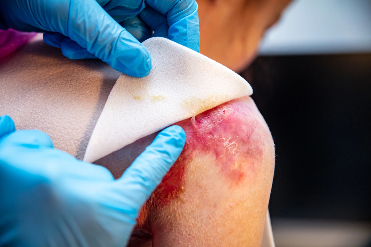
Skin biopsy and immunofluorescence mapping
One of the primary diagnostic tools for EB is a skin biopsy, typically taken from the edge of a fresh blister. The sample is then subjected to immunofluorescence mapping, a technique where fluorescently labelled antibodies are used to detect specific structural proteins in the skin. This process reveals which proteins are missing, reduced, or malfunctioning and identifies the layer of skin where the separation occurs.
Immunofluorescence mapping is an efficient method for diagnosing EB, as it helps clinicians differentiate EB from other blistering conditions and clarifies which specific subtype the patient has. It also informs genetic testing strategies, aiding clinicians in determining which genes to focus on. For many healthcare providers, this test, alongside genetic analysis, is a cornerstone of congenital epidermolysis bullosa diagnosis.
Electron microscopy
Although less commonly used than immunofluorescence mapping, electron microscopy provides highly detailed images of the skin’s cellular structure. This technique can identify the exact point where the skin layers separate by producing high-resolution images of the dermo-epidermal junction. Transmission electron microscopy (TEM) can be particularly useful in cases where immunofluorescence results are inconclusive, adding important details to support the diagnosis. However, access to this advanced technique may be limited by the availability of specialized equipment and expertise.
Genetic testing
For the most accurate diagnosis, healthcare providers often combine the results of immunofluorescence mapping and genetic testing. The first method reveals which structural proteins are present or missing in the skin tissue, while the second identifies the exact mutation responsible for the condition. When used together, these tests help reduce diagnostic uncertainty and provide crucial information to produce tailored plans for treatment of congenital epidermolysis bullosa.
Some medical centres prioritize genetic panels as the first-line test for EB because they are highly accurate and non-invasive. This approach can shorten the time to a definitive diagnosis and may reduce the need for a skin biopsy. In facilities with advanced laboratory capabilities, using genetic testing as the initial step is becoming more common.
References
- Siañez González C, Pezoa Jares R, Salas Alanís JC. Congenital epidermolysis bullosa: a review. Apunts Med Esport [Internet]. 2009 [cited 2025 Dec 15]. Available from: https://www.apunts.org/en-download-pdf-S1578219009705542
- Immunofluorescence mapping for diagnosis of congenital epidermolysis bullosa. Actas Dermosifiliogr [Internet]. [cited 2025 Dec 15]. Available from: https://www.actasdermo.org/es-immunofluorescence-mapping-for-diagnosis-congenital-articulo-S1578219…
- Genetic diagnosis of epidermolysis bullosa: recommendations from an expert Spanish research group. Actas Dermosifiliogr [Internet]. [cited 2025 Dec 15]. Available from: https://www.actasdermo.org/en-genetic-diagnosis-epidermolysis-bullosa-recommendations-articulo-S157…
- Has C, et al. Clinical practice guidelines for laboratory diagnosis of inherited epidermolysis bullosa. Br J Dermatol [Internet]. 2019 [cited 2025 Dec 15]. Available from: https://pmc.ncbi.nlm.nih.gov/articles/PMC7064925/ PMC