Onychomycosis diagnosis
What are the main methods of diagnosis for onychomycosis?
Accurate diagnosis of onychomycosis (fungal nail infection) is essential, as several other nail conditions—such as psoriasis, traumatic changes or lichen planus—can mimic its appearance. Correct identification helps ensure that treatment is appropriate and avoids unnecessary use of antifungal medicines.
In order to correctly diagnose onychomycosis, healthcare professionals may resort to:
A potassium hydroxide (KOH) preparation
A small sample of nail material or debris taken from beneath the nail is treated with potassium hydroxide and examined under a microscope. This technique allows clinicians to look directly for fungal structures. It is quick, inexpensive, and widely available, though its sensitivity can vary and false-negative results do occur.
Fungal culture
A sample can be placed on a specialized medium to encourage fungal growth. This technique enables us to identify the exact species involved, which can guide treatment decisions. However, it usually requires several weeks for results and may be less sensitive if the fungi are not viable or if the patient has recently used antifungal products.
Histopathology (sectioning of the nail plate)
A nail clipping can be preserved, thinly sectioned, and stained to highlight fungal elements within the nail itself. This method often detects infection more reliably than fungal cultures and helps confirm the presence of fungi in situ.
Polymerase chain reaction (PCR) and other molecular tests
Modern molecular techniques detect fungal DNA directly from nail samples. These tests tend to be more sensitive and provide quicker results than fungal cultures. They can identify a wide range of fungi, including dermatophytes, yeasts and non-dermatophyte moulds, and are increasingly used in specialist settings.
Dermoscopy and reflectance confocal microscopy
Non-invasive imaging tools may help support a clinical diagnosis. Dermoscopy provides a magnified view of the nail surface, while confocal microscopy allows real-time imaging of deeper layers. Both can reveal patterns typically associated with fungal infection, although they cannot replace laboratory confirmation.
If you attend a medical appointment because of a suspected fungal nail infection, you can expect a structured assessment that usually includes the following steps:
Clinical examination
The clinician will examine the appearance of the affected nail or nails, noting changes such as discolouration, thickening, separation of the nail from the nail bed, or build-up of debris. The pattern of involvement—for example, affecting the distal edge, the surface, or the entire nail—may help narrow down the diagnosis.
Sample collection
If onychomycosis is considered likely, a sample will be obtained. This may involve trimming the nail, scraping material from underneath, or gently debriding the affected area. Obtaining material from deeper, more active parts of the infection improves the reliability of laboratory tests.
Laboratory analysis
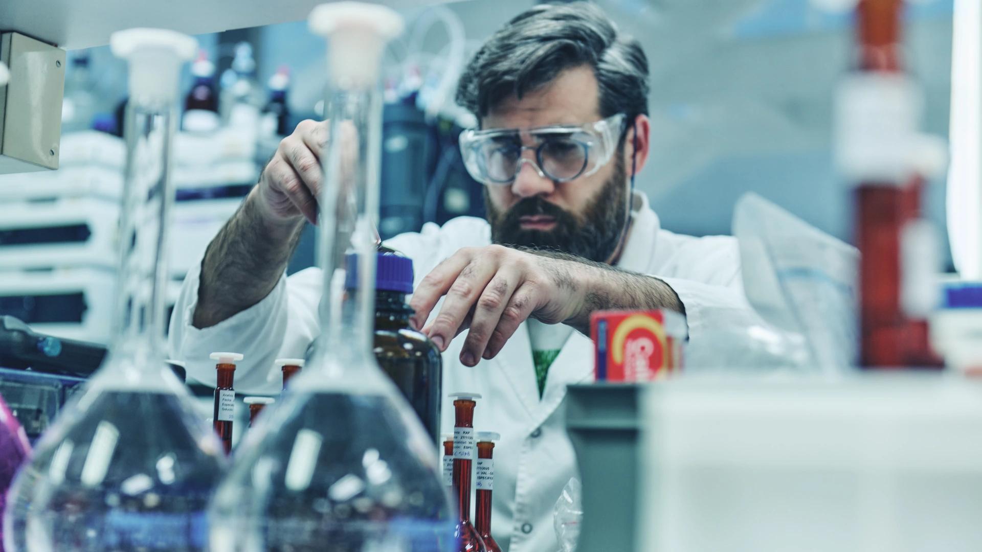
The collected material is analysed using one or more of the available techniques. Microscopy with KOH offers quick initial information, while fungal cultures help determine the fungal species. Histopathology and molecular testing provide additional clarity, especially in cases where the diagnosis is uncertain or previous tests have been inconclusive. Each method contributes a different type of evidence.
Diagnosis of onychomycosis and discussion
Once the laboratory results are available, after a few days or several weeks, the clinician will review the findings with you. This discussion usually covers whether a fungal infection has been confirmed, which organism is responsible, and which treatment options for onychomycosis are most suitable. Great care should be taken when prescribing systemic antifungal medicines, so confirmation of infection is recommended before starting them.
Accurate diagnosis is the cornerstone of effective management of onychomycosis. Clinical examination can strongly suggest the condition, but laboratory confirmation helps us distinguish it from other nail disorders, identify the causative organism, and proposethe most appropriate treatment. By ensuring that the diagnosis is correct from the outset, patients receive targeted care and avoid unnecessary or ineffective therapies.
References
- Verma S, Daniels I. Diagnosis of Onychomycosis: From Conventional Techniques and Emerging Molecular Assays. Frontiers in Medicine [Internet]. 2021 [cited 2025 Nov 18]; 8:637216. Available at: https://www.frontiersin.org/journals/medicine/articles/10.3389/fmed.2021.637216/full
- Hay RJ, Baran R. Onychomycosis: An updated review. PM CID: PMC7509699 [Internet]. 2020 [cited 2025 Nov 18]; 10:742. Available at: https://pmc.ncbi.nlm.nih.gov/articles/PMC7509699/
- Lipner SR, Scher RK. Onychomycosis: Current Trends in Diagnosis and Treatment. American Family Physician [Internet]. 2013 [cited 2025 Nov 18]; 87(7):662-670. Available at: https://www.aafp.org/pubs/afp/issues/2013/1201/p762.html/
- Miller J. Onychomycosis: Rapid Evidence Review. American Family Physician [Internet]. 2021 [cited 2025 Nov 18]; 103(3):145-152. Available at: https://www.aafp.org/pubs/afp/issues/2021/1000/p359.html